Subscribe to Receive My HealtheVet Updates
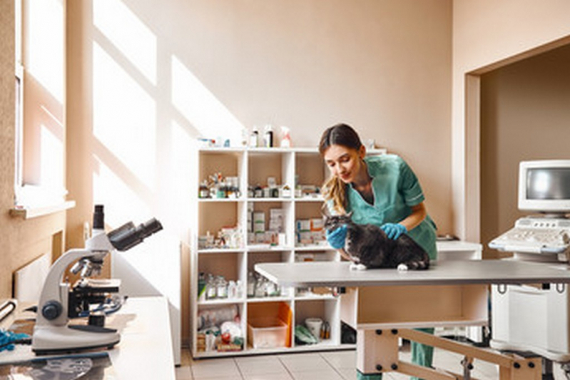
In most trendy x-ray machines, the technique chart is built into the machine. The operator want solely enter the species, physique part, and thickness, and the machine mechanically sets the approach. This is convenient and reduces mistakes in approach, however the settings might need to be altered to suit the precise equipment, film-screen (detector) pace, and viewer’s preferences (eg, contrast level). Ultrasonography (commonly referred to as ultrasound) is the second mostly used imaging procedure in veterinary practices. It makes use of sound waves to create pictures of body buildings primarily based on the sample of echoes reflected from the tissues and organs.
Veterinary Radiology
Veterinary radiologists are vets who full veterinary faculty and then go on to do a radiology residency for several years. IV and intra-arterial contrast agents are typically iodine primarily based and improve the opacity of the blood, making vascular constructions seen. Iodinated distinction agents are cleared primarily by the kidneys, making the amassing system of the urinary tract seen. Orally administered brokers, primarily barium sulfate–based compounds, outline the mucosa and lumen of the GI tract. Intrathecal distinction agents are additionally iodine primarily based and allow analysis of the spinal wire and meninges.
The emitted waves are then transformed into pictures which might be displayed on a computer display. Sequential examination of slices by way of the physique is completed in the identical way as for computed tomography. Because the process is quite prolonged and the animal should not transfer throughout the process, general anesthesia is used generally. In this procedure, the animal is placed on a motorized bed inside a CT scanner, which takes a collection of x-rays from totally different angles.
Differences Between Human and Veterinary Medicine and X-Rays
Monitoring of exposure additionally supplies evidence of proper adherence to radiation security requirements if questions arise as to whether an employee’s medical condition could be related to radiation publicity. Proper positioning is also essential to maximize the diagnostic content material of the x-ray examination. In many instances, improper positioning or radiographic examination may find yourself in a misdiagnosis or inability to understand main lesions. Both proper and left lateral recumbent radiographs are beneficial in dogs and cats. This is completed as a result of positioning of the animal on its aspect ends in fast relocation of fluids to and atelectasis of the downside lung.
Canine Medical Imaging, Ultrasound, X-rays, https://Postheaven.Net Radiographs
Automatic publicity management (AEC) is a system during which the operator units the kVp and mA, and the machine terminates the publicity on the applicable time. If used correctly, this technique leads to nearly similar picture exposures between animals. However, acceptable kV settings are wanted, and constant animal positioning is critical. Identical positioning between animals is required to attain identical images. Placing the heart or lungs over the AEC sensor ends in radically completely different radiographs. AEC might be most effective when massive numbers of pictures are being done of the same anatomic space by the same personnel. AEC is typically not used in most veterinary applications due to the broad variation in physique sizes and conformation of dogs.
 Además de esto, esta herramienta jamás debe usarse para cortar ninguna anatomía del tolerante capturada por la exposición inicial y la reconstrucción. Las radiografías se han empleado durante muchas décadas para hacer imágenes llamadas radiografías (imágenes en negro, blanco y gris). La radiografía se encuentra dentro de las herramientas diagnósticas más muchas veces utilizadas en las clínicas veterinarias. Las imágenes de rayos X (radiografías) se generan con los mismos procesos empleados en la medicina humana, salvo que el equipo está dimensionado para su uso con perros, gatos y otros pequeños animales. Los equipos portátiles pueden usarse en una clínica de enormes animales que trate caballos y otros enormes animales. Aunque el trámite es indoloro, en algunos casos las mascotas se sedan para reducir la ansiedad y el estrés socios con el procedimiento, para ubicar al animal y para ayudarlo a permanecer inmovil mientras que se toman las imágenes. Los animales han de estar adecuadamente sujetos y posicionados para obtener imágenes radiográficas laboratorio de analises clinicas veterinarias calidad.


